When researchers in ╣¹¢┤╩ËãÁÔÇÖs (NCIL) designed a huge, two-year reading comprehension study involving approximately 100 children from grades two and three, they faced a major challenge. They had to decipher the intricacies of neuroplasticity ÔÇö how the brain rewires itself to do new things ÔÇö during the formative stages of reading while simultaneously shepherding children through the intimidating world of Magnetic Resonance Imaging (MRI) scans.
ÔÇ£Having children come in for a (voluntary) MRI scan is loaded with a high risk of attrition, meaning the risk is great that they won't finish the scan, or they might drop out of the study entirely,ÔÇØ says Dr. Cindy Hamon-Hill, researcher and lab manager with NCIL, a group led by Dr. Aaron Newman in the Department of Psychology and Neuroscience.
Most children have never had an MRI (basically, a scan used to capture high-resolution images of internal anatomy), let alone a functional MRI on the brain, while doing a task. To avoid blurry images and to obtain usable data, subjects are required to stay still for approximately thirty minutes ÔÇö a struggle for little children. (In pediatric studies involving MRI scans, a 50 per cent success rate is considered good; often, most of the data is deemed unusable.)
To mitigate the challenge, the researchers prescribe some play homework for all study participants in advance of their scan: the children are asked to crawl inside a nylon play tunnel supplied by the lab and lay down, playing the ÔÇ£statue gameÔÇØ while a parent times how long they can keep still. The children are encouraged to play the game several times a week and to work at increasing their ÔÇ£lying-still-timeÔÇØ with each session.┬á
A light-bulb moment
While the statue game is helpful, researchers wondered if it would be enough preparation for an MRI. To get the best possible data, the lab needed something to further prepare the children for the MRI experience. While the IWK Health Centre possesses a mock MRI, its limited availability made it impossible to use for their study.
 In a lightbulb moment, an idea emerged ÔÇö the lab would construct its own mock MRI specifically designed to train the children for the experience.
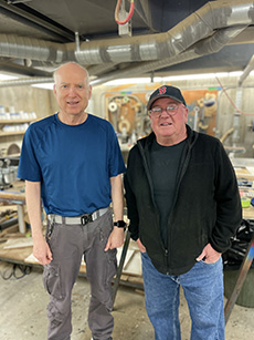
The lab approached technicians Chris Wright and Mark Merrimen (shown left and right, respectively) with their idea, and the two men took on the unconventional project with gusto last winter. ÔÇ£It was a rather large project, with lots of imaginative and inventive design work done by Mark and Chris,ÔÇØ says Dr. Hamon-Hill.┬á
Merrimen was new to the department at the time, having just come from 25 years in Oceanography and a long career in aircraft and boat building. The lab showed him a picture of the IWKÔÇÖs mock MRI and asked if he could build something similar. He didnÔÇÖt hesitate.
ÔÇ£I thought, ÔÇÿYeah, we can do that,ÔÇØ says Merrimen, who obtained some general dimensions and got to work.┬á
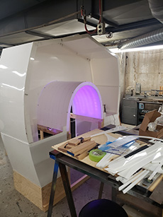
He researched what kinds of materials they could use that could be manipulated and shaped, designing and solving problems as he went along. ÔÇ£It was kind of nice in a way because I could employ any type of material I wanted, any type of construction,ÔÇØ he says. ÔÇ£I basically built it out of a frame-and-skin construction with high-density polyethylene framing."
Merrimen used wood for the frame to save on costs.
Lights, sounds, action
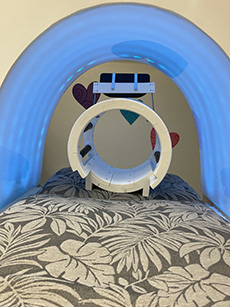
Meanwhile, Wright used a 3-D printer to build a mock ÔÇ£head coilÔÇØ ÔÇö the part that the head rests in inside the MRI. He also came up with the idea to include some programmable coloured Bluetooth LED lighting in the tunnel to further simulate an electronic experience.┬á
 ÔÇ£I was thinking maybe it would get the kids relaxed . . . having a (coloured light) pattern that they would enjoy,ÔÇØ he says.
Shown right: The mock MRI under construction. (Mark Marrimen image)
He also helped set up the speakers for some MRI sound effects the lab put together that were sourced from real-life MRI audio, which is typically loud and full of knocking, creaking, and sawing noises.
The whole project was achieved on a next-to-nothing budget ÔÇö a testament to the techniciansÔÇÖ resourcefulness and innovative thinking.
Several months of work culminated in April, when they unveiled the labÔÇÖs new true-to-size mock MRI. Complete with a plywood bed, mattress, neck brace, and authentic MRI sound effects, it exceeded expectations.
ÔÇ£I didn't think it would be quite this elaborate, but they built it to the same proportions, depth-wise and width, and with materials that would be solid so that we can move it around and use it again and try to make it as safe as possible,ÔÇØ says Dr. Hamon-Hill.
A community resource
 So far, 12 children in the study have done practice sessions using the labÔÇÖs Mock MRI, and only two of those were not able to make it through the MRI at the IWK ÔÇö a much better result than the typical rates. Of the two that didnÔÇÖt go through with the MRI, one of them is keen to try the MRI again, having spent some additional time training in the mock MRI.
The labÔÇÖs two-year study continues, but Dr. Hamon-Hill wants other labs at ╣¹¢┤╩ËãÁ to know about the mock MRI and its availability for research.
ÔÇ£We have so many people working with children for clinical reasons If they have to go for MRIs, they could come here for practice sessions,ÔÇØ she says.
Interested? Please contact Cindy Hamon-Hill with the NeuroCognitive Imaging Lab (NCIL). 

